
Comparing macular degeneration vs glaucoma can feel confusing because both can affect vision, both become more common with age, and both often start quietly. If you’re trying to protect your long-term sight—especially if you’ve noticed changes like blur, trouble reading, glare, or reduced side vision—knowing how these conditions differ can help you ask better questions at your next visit and act sooner when symptoms show up. As fall routines settle in, it’s a good time to reassess eye health habits and make sure your screenings match your risk factors.
This guide breaks down what each condition affects, typical symptoms, how they’re usually diagnosed, and how follow-up care often looks. For a broader overview of evaluation and treatment options, you can also review Georgia Eye’s ophthalmology services.
If you’re looking for local care, start with an eye doctor consultation for macular degeneration vs glaucoma in Athens, GA to discuss symptoms, risk factors, and the right testing schedule.
Bottom Line Upfront: macular degeneration vs glaucoma
- Macular degeneration primarily affects central vision (the “fine detail” you use for reading and recognizing faces).
- Glaucoma more often affects peripheral (side) vision first and can progress without obvious early symptoms.
- Both conditions can be present at the same time, so “one diagnosis” doesn’t automatically rule out the other.
- Diagnosis typically relies on in-office testing (imaging and functional vision tests), not symptoms alone. This article is educational and not a diagnosis.
- Consistent monitoring is often more protective than waiting for noticeable vision loss.
How These Two Conditions Differ in the Eye
The simplest way to compare macular degeneration vs glaucoma is to focus on what part of the visual system is affected:
- Macular degeneration (often age-related) involves changes in the macula, the central part of the retina responsible for sharp, straight-ahead vision.
- Glaucoma involves damage to the optic nerve (the “cable” carrying visual information to the brain). It’s commonly associated with eye pressure, but pressure is only one part of the picture.
Because the macula and the optic nerve serve different roles, the day-to-day visual complaints can look different—even if both conditions fall under the same umbrella of chronic eye disease. Symptoms can overlap with other issues (like dry eye, cataracts, or prescription changes), so an eye exam is typically the most reliable way to sort out the cause.
Side-by-Side Comparison Table (Symptoms, Tests, Daily Impact)
| Comparison point | Macular degeneration | Glaucoma |
|---|---|---|
| Vision affected first (typical pattern) | Central detail vision (reading, faces) | Peripheral vision (side vision); central vision later |
| Common early clues | Wavy lines, missing spots in the center, more trouble in dim light | Often none at first; subtle bumping into objects or trouble in low light for some |
| How it’s often detected | Retinal exam + retinal imaging; central vision testing | Eye pressure check + optic nerve evaluation + visual field testing |
| Daily-life impact | Reading, driving signs, recognizing faces, fine work | Mobility, driving awareness, navigating stairs/curbs, side awareness |
| Monitoring focus | Retinal changes and central function over time | Optic nerve stability and field changes over time |
The Real-World Cost of Waiting (Time, Safety, and Follow-Up)
With macular degeneration vs glaucoma, the biggest “cost” is often lost opportunity for early detection and structured monitoring. Practical implications can include:
- Safety: Reduced side vision can increase fall risk and driving hazards; reduced central vision can affect reading labels, medication instructions, and recognizing faces.
- Time: Later-stage care can mean more frequent visits and more complex decision-making.
- Budget/value: Ongoing monitoring can feel like a hassle, but it may be more cost-effective than reacting to sudden functional changes and needing more intensive support.
If you’re already seeing changes, don’t self-diagnose based on online symptom lists. A comprehensive eye exam is typically the most reliable way to separate common causes of blur (like dry eye or a glasses change) from retinal or optic nerve concerns.
Common Missteps That Delay Diagnosis (Checklist)
- Assuming blur always equals “needs new glasses” — refraction matters, but retinal/nerve problems can also change clarity.
- Skipping follow-ups after a “borderline” result — trend over time is often what clarifies risk.
- Relying on symptoms alone — glaucoma in particular can progress quietly until side vision loss is noticeable.
- Not bringing a medication list — some medicines and health conditions can influence eye findings and testing choices.
- Ignoring one-eye changes — differences between eyes can be a useful clue and should be evaluated.
A Smart Prep Plan for Your Next Eye Visit (Checklist)
- Write down specific vision problems (example: “straight lines look wavy,” “missing spots,” “night driving feels harder,” “bumping into things on one side”).
- Bring your current glasses/contacts info so the team can separate prescription issues from health issues.
- Ask what tests you’re having (imaging, visual field testing, pressure checks) and what each test is meant to answer.
- Confirm your follow-up interval and what changes should prompt an earlier visit.
- Plan for dilation when advised—it can affect driving and work for a few hours.
Professional Insight: The Symptom Pattern That Helps Most
Clinicians often find pattern-based descriptions more actionable than vague ones—“my center is distorted” versus “my vision is worse,” or “I’m missing things off to the side” versus “I’m clumsy lately.” Clear details (which eye, when it started, whether it’s constant, and what tasks are affected) can help your eye doctor decide which tests to run first and how to compare results over time. This is general guidance, not a diagnosis.
When to Seek an Eye Doctor’s Evaluation
Schedule an evaluation if you notice any of the following:
- New central distortion (straight lines bending, missing/blurred spots in the center)
- New peripheral vision concerns (trouble noticing objects on one side, bumping into doorframes)
- Rapid change in vision in one or both eyes
- Difficulty with driving safety (especially lane awareness or reading signs)
- Family history concerns you haven’t discussed with an eye doctor yet
If you believe you’re having an emergency, seek immediate medical care. Routine ophthalmology clinics generally operate during standard office hours.
Common Questions People Ask
Can you have both conditions at the same time?
Yes. The retina (macula) and optic nerve are different structures, so it’s possible to have findings related to both. An exam and targeted testing help clarify what’s happening in each eye.
Which one affects reading more?
Central-vision problems tend to interfere with reading more directly. Side-vision loss can also make reading harder (tracking lines, scanning), but the pattern is often different.
Do symptoms always show up early?
No. Some people notice changes early, while others don’t—especially if only one eye is affected at first or if changes are gradual.
What should I bring to my appointment?
Bring your glasses or contact lens information, a current medication list, and a short note describing when symptoms started and what tasks are harder (reading, night driving, stairs, screens). If you’ve had prior eye records or imaging, bring those too if available.
Moving Forward
Comparing macular degeneration vs glaucoma comes down to where the damage occurs and how it shows up in daily life—central detail versus side awareness. The most practical next step is a structured exam and a monitoring plan that matches your risk and symptoms. If you’re unsure what you’re experiencing, describing the pattern clearly is often more useful than trying to label the condition yourself.
Get Your Free Quote
For a medically appropriate next step, schedule an eye exam/consultation and bring your medication list. Ask whether you may need dilation, retinal imaging (like OCT), or visual field testing so you can plan your day and transportation.
What Tests Help Differentiate Macular Degeneration vs Glaucoma?
Symptoms can suggest a pattern, but testing is typically needed to tell macular degeneration vs glaucoma apart (and to check for other causes). Here are common tests and what they help evaluate:
- Dilated fundus exam: Allows a direct look at the retina and optic nerve. It can help identify macular changes (like drusen or pigment changes) and optic nerve findings that raise concern for glaucoma.
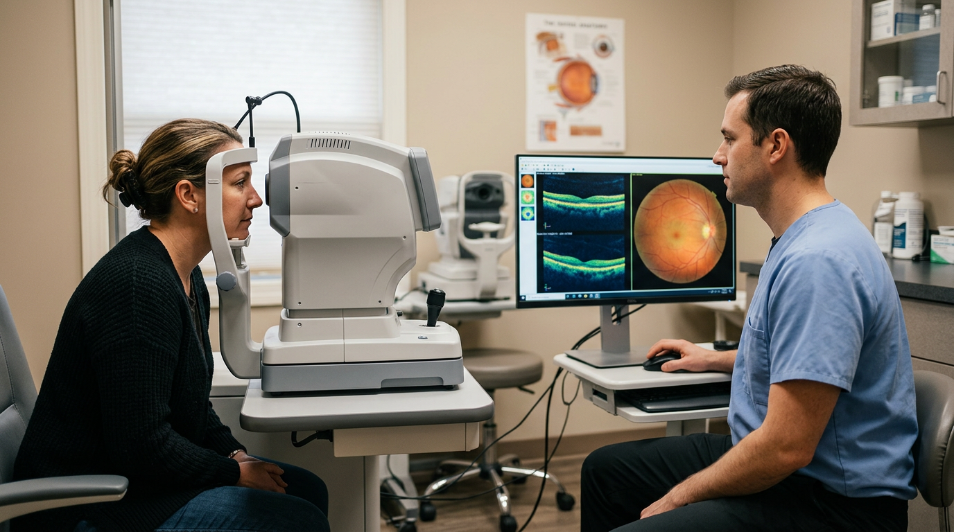
- OCT (Optical Coherence Tomography): Imaging that measures retinal layers. It can help assess the macula for swelling/structural changes and measure the retinal nerve fiber layer (RNFL) and ganglion cell complex for glaucoma-related thinning.
- Visual field test (HVF/perimetry): Measures functional side vision and can detect patterns of loss that are typical for glaucoma. It’s also useful for tracking change over time.
- Tonometry (eye pressure check): Measures intraocular pressure. Higher pressure can increase glaucoma risk, but normal pressure does not rule glaucoma out.
- Pachymetry (corneal thickness): Measures corneal thickness, which can affect how eye pressure readings are interpreted and can help refine glaucoma risk assessment.
- Gonioscopy: Examines the drainage angle of the eye to help determine whether the angle is open or narrow/closed—important for classifying glaucoma risk and guiding management.
- Amsler grid discussion/testing: A simple way to screen for central distortion (wavy lines or missing areas) that can be associated with macular disease. It doesn’t replace an exam, but it can help you monitor changes between visits.
How Often Are Follow-Ups Typically Scheduled?
Follow-up timing varies by diagnosis, severity, and risk factors. These are general ranges many clinicians use as a starting point (your plan may differ):
- Glaucoma suspect / borderline findings: often every 6–12 months, sometimes sooner if testing is being repeated to confirm a baseline.
- Diagnosed glaucoma (stable): often every 3–6 months, with periodic visual fields and OCT to watch for progression.
- Macular degeneration (early/stable): often every 6–12 months, with earlier visits if new distortion or central changes appear.
- Macular degeneration with new or worsening symptoms: evaluation is often recommended promptly rather than waiting for the next routine check.